Instrumentation & Services
Instrumentation
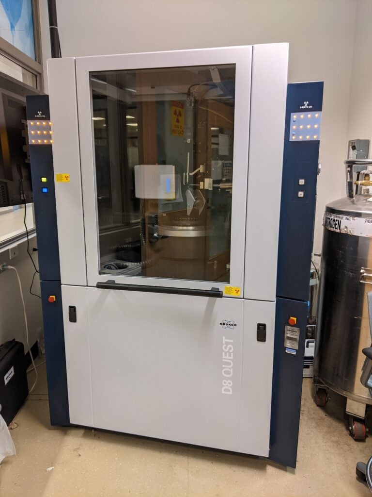
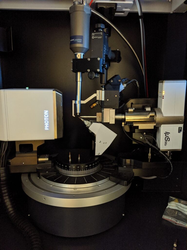
Bruker D8 Quest – 234A Regents Hall
- Mo microfocus sealed tube
- Photon 100 CMOS detector
- Oxford Cryostream 700
Uses: variable temperature single crystal X-ray diffraction experiments
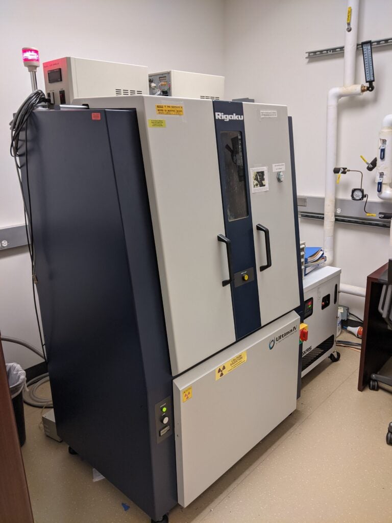
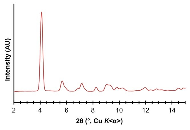
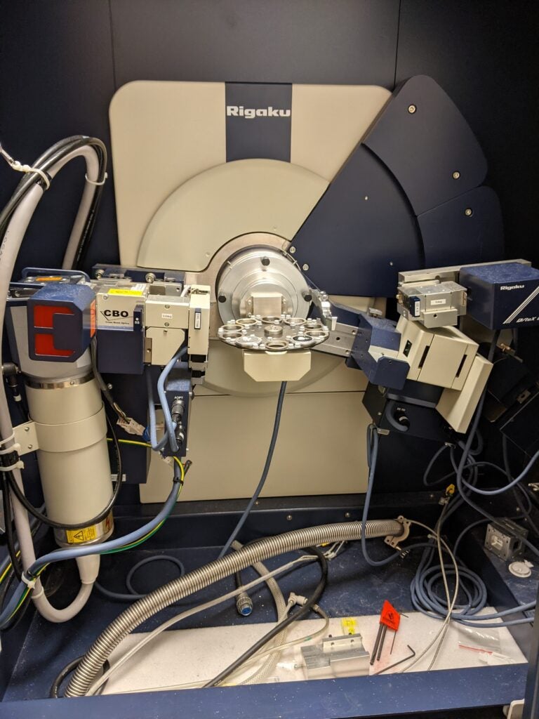
Rigaku UltimaIV – 240 Regents Hall
- Cu fine-focus sealed tube
- Dual detector instrument:
- DTeX Ultra
- Scintillation Counter
- Zero background Si wafer sample holders
- Various attachments
Uses: powder X-ray diffraction experiments in Bragg-Brentano geometry
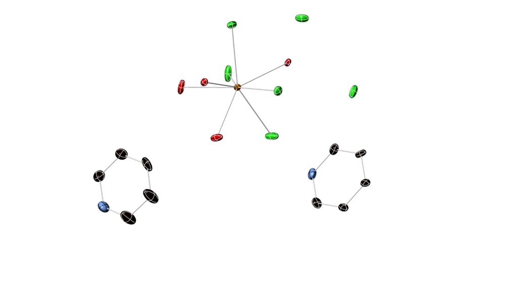
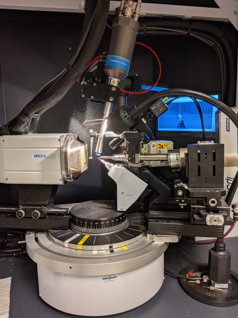
Bruker APEX DUO – 127 Basic Science Building
- Dual source instrument:
- Mo fine-focus sealed tube
- Cu microfocus sealed tube
- Apex II CCD detector
- Oxford Cryostream 700
Uses: variable temperature single crystal and powder X-ray diffraction experiments
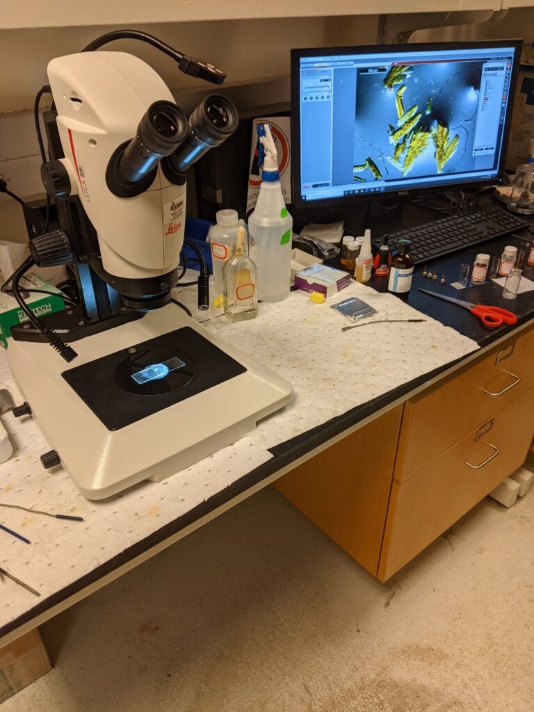
Leica Stereozoom S9i Microscope – 127 Basic Science Building
- Up to 55x magnification
- Polarizing analyzer
- Integrated 10 megapixel camera
- Computer running Leica Application Suite:
- save images of crystals
- measure crystal size
- annotate saved images
Services
Georgetown University researchers who intend to incorporate X-ray diffraction as a regular tool in their analytical toolbox are strongly encouraged to become fully trained instrument users. Georgetown researchers who do not intend to regularly use the X-ray facilities can simply contact Dr. Bertke to discuss experiments and submit samples. Researchers outside of Georgetown University should contact Dr. Bertke to discuss experimental needs and pricing.
- Training on all facility instruments
- Training and instruction on single crystal refinement software (ShelXle)
- Help with non-trivial structure refinements
- Full or partial service single crystal data collection and processing
- Powder diffraction data collection
- Crystal growth advice
- Advice on experiment selection
- Manuscript prep and editing